Podemos decir que la familia (Erebidae) es una de las mas grandes, con un aproximado de 45 mil géneros alrededor del mundo. En Area de Conservación Guanacaste (ACG) tenemos un aproximado de unos 400 géneros con 1500 especies, estos registros se han dado a lo largo de 35 años del proyecto de buscar y criar larvas en diferentes estaciones del ACG por los Gusaneros, sin contemplar el resto del país.
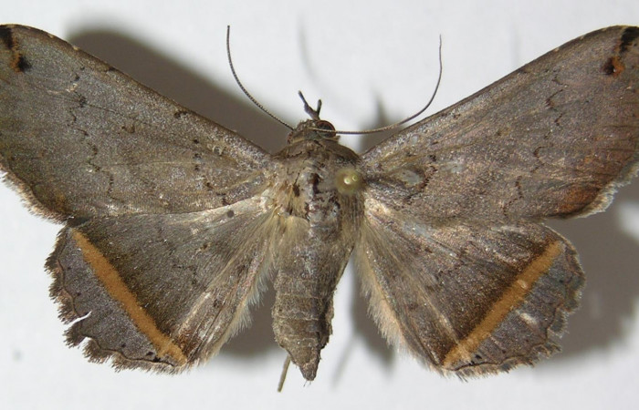
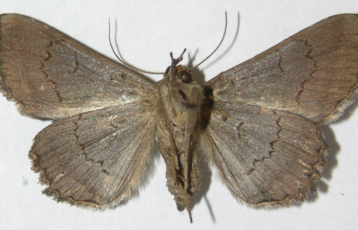
Adulto Lesmone formularis (Erebidae), de hábitos nocturnos, tamaño medio, su envergadura alar es de 35 mm.
 Figura 1. Vista dorsal Lesmone formularis (Erebidae), envergadura alar 35 mm. Voucher: 09-SRNP-76232-DHJ522554.jpg.
 Figura 2. Vista dorsal Lesmone formularis (Erebidae), envergadura alar 35 mm. Voucher: 09-SRNP-76232-DHJ522555.jpg.
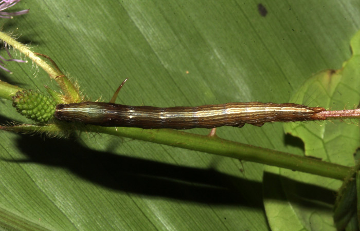
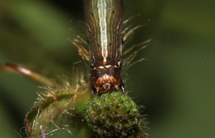
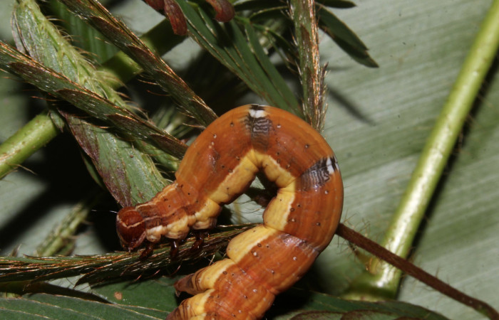
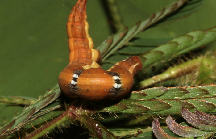
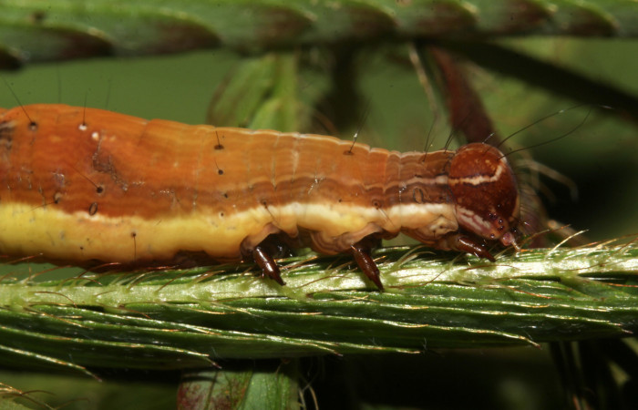
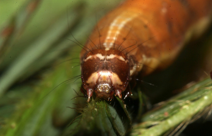
Larva Lesmone formularis (Erebidae) en tercer estadío:
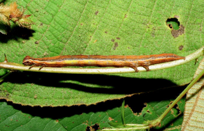
Larva de color café, líneas finas de color crema en todo el cuerpo, línea dorsal amarilla muy visible, tiene pelitos muy finos negros en partes de los segmentos del cuerpo.
Larva se camufla muy bien en los bejucos y enredaderas que se forman en los charrales.
 Figura 3. Larva de Lesmone formularis (Erebidae). Vista lateral, penultimo estadío 32 mm. Voucher: 18-SRNP-32106-DHJ748345.jpg.
 Figura 4. Larva de Lesmone formularis (Erebidae). Vista dorsal, penultimo estadío 32 mm. Voucher: 18-SRNP-32106-DHJ748346.jpg.
 Figura 5. Cabeza de frente Lesmone formularis (Erebidae), penultimo estadío 32 mm. Voucher: 18-SRNP-32106-DHJ748350.jpg.
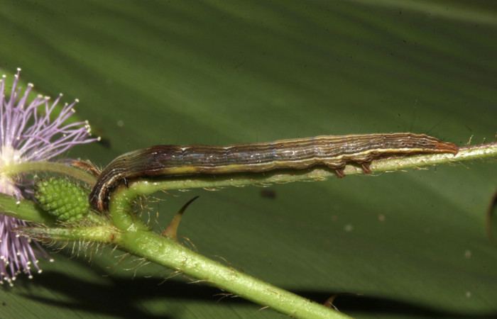
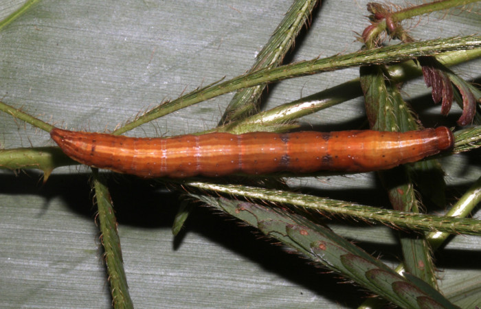
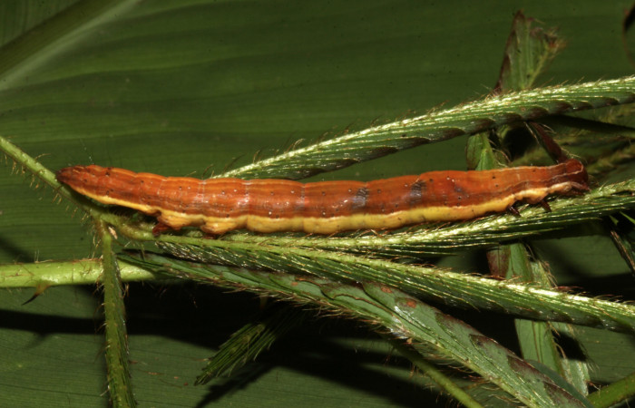
Larva Lesmone formularis (Erebidae) en último estadio:
Cuando la larva de Lesmone formularis (Erebidae) se encuentra estirada se le pueden ver en la parte dorsal cuatro puntos negros y en los costados dos (Fig. 6-7).
 Figura 6. Larva de Lesmone formularis (Erebidae). Vista dorsal, último estadío 49 mm. Voucher: 18-SRNP-32105-DHJ748335.jpg.
 Figura 7. Larva de Lesmone formularis (Erebidae). Vista lateral, último estadío 49 mm. Voucher: 18-SRNP-32105-DHJ748336.jpg.
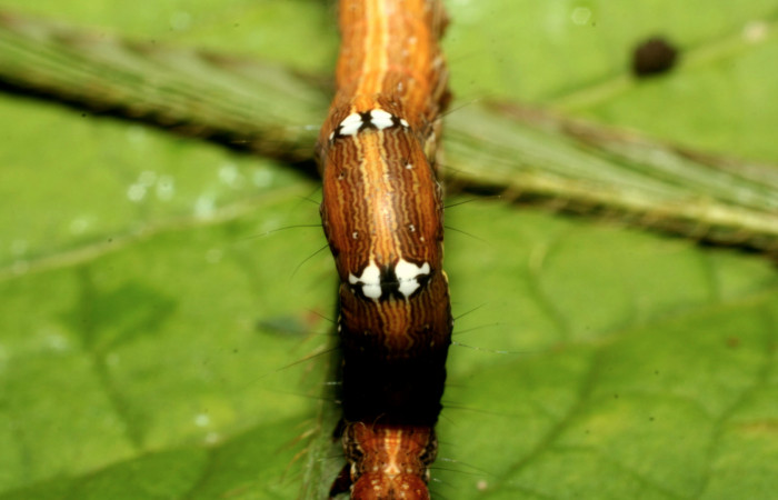
Cuando la larva se mueve o empieza a caminar se le pueden mirar dos manchas blancas en la parte dorsal con un punto negro a la par (Fig. 8-9).
 Figura 8. Larva de Lesmone formularis (Erebidae). Lateral torax, último estadío 49 mm. Voucher: 18-SRNP-32105-DHJ748337.jpg.
 Figura 9. Larva de Lesmone formularis (Erebidae). Dorsal torax, último estadío 49 mm. Voucher: 18-SRNP-32105-DHJ748338.jpg.
 Figura 10. Larva de Lesmone formularis (Erebidae). Cabeza de lado, último estadío 49 mm. Voucher: 18-SRNP-32105-DHJ748342.jpg.
 Figura 11. Larva de Lesmone formularis (Erebidae). Cabeza de frente, último estadío 49 mm. Voucher: 18-SRNP-32105-DHJ748341.jpg.
Capullo de Lesmone formularis (Erebidae):
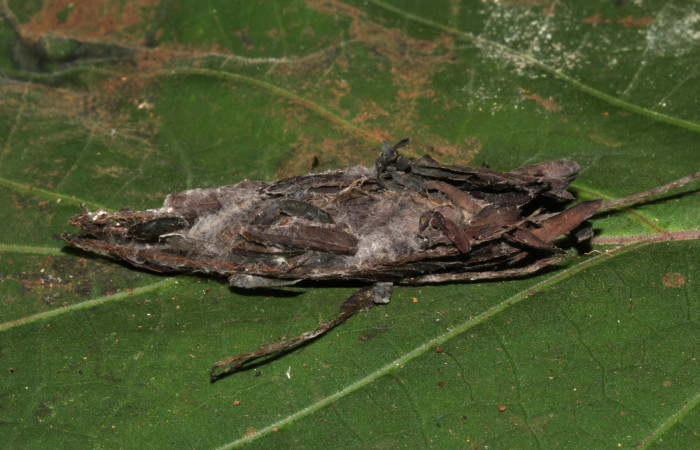
Cuando la larva deja de comer, empieza el proceso de prepupa, el cual es la transformación para formar la pupa. Dependiendo de la familia de larva, no todas hacen nidos o capullos, en este caso la larva empieza a hacer un nido hecho con pedazos de hojas secas que se pone encima del capullo que va tejiendo con su seda, (Fig. 12), en el cual ya terminado y bien cerrado, empieza la prepupa a moverse para quitarse la cutícula o piel que usó en el último estadío para formar la pupa.
 Figura 12. Capullo de Lesmone formularis (Erebidae). Capullo 50 mm. Foto 21/diciembre/2018. Voucher: 18-SRNP-32105-DHJ748503.jpg.
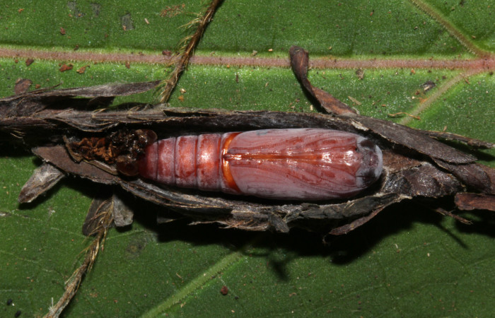
Pupa de Lesmone formularis (Erebidae):
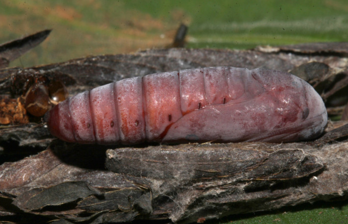
Pupa de color rojo, 20 mm de longitud, se le puede ver los espiráculos en la parte lateral (Fig. 13).
 Figura 13. Pupa lateral de Lesmone formularis (Erebidae). Pupa 20 mm. Foto: 21/diciembre/2018. Voucher: 18-SRNP-32105-DHJ748506.jpg.
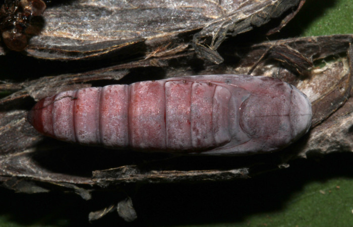
 Figura 14. Pupa dorsal de Lesmone formularis (Erebidae). Pupa 20 mm. Foto: 21/diciembre/2018. Voucher: 18-SRNP-32105-DHJ748507.jpg.
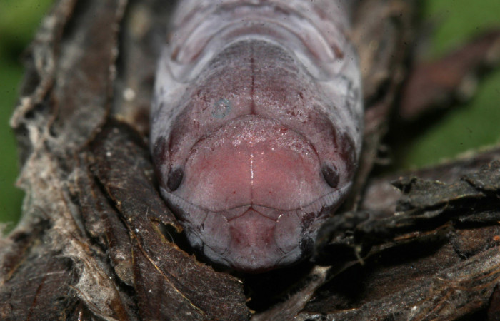
 Figura 15. Pupa ventral de Lesmone formularis (Erebidae). Pupa 20 mm. Foto: 21/diciembre/2018. Voucher: 18-SRNP-32105-DHJ748505.jpg.
 Figura 16. Pupa de frente Lesmone formularis (Erebidae). Pupa 20 mm. Foto: 21/diciembre/2018. Voucher: 18-SRNP-32105-DHJ748511.jpg.
Parásitos de Lesmone formularis (Erebidae):
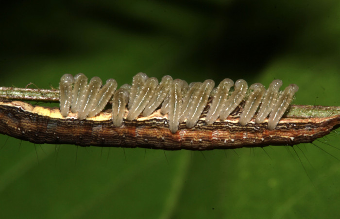
La avispa de Braconidae cuando esta volando, encuentra la larva de mariposa donde va inyectar su huevo dentro la larva, las larvitas en primer estadío sale del huevo y empieza comiendo la sangre de la larva. Cuando la larva de la avispa está completamente desarrollada, rompe la cutícula (piel) de la larva y sale por el hueco. Afuera, hace su capullo de seda que puede quedar pegada a la cutícula o pegados en ramitas. La larva de la mariposa puede seguir caminando pero pronto muere.
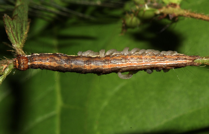
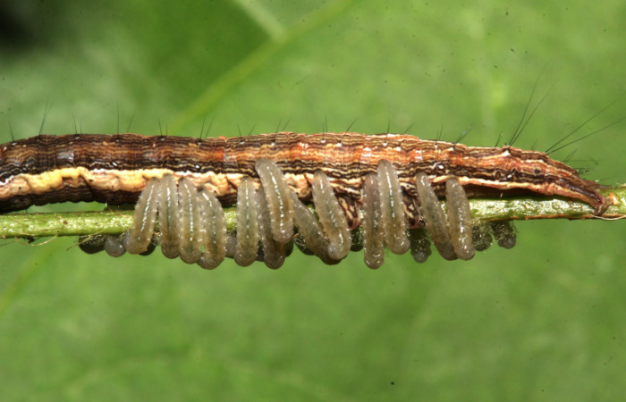
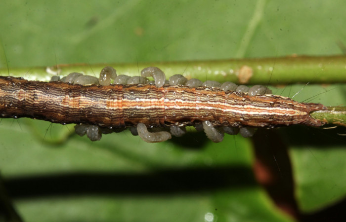
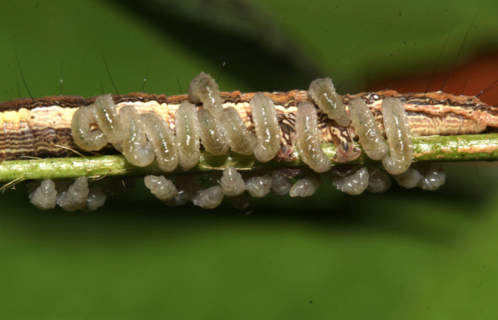
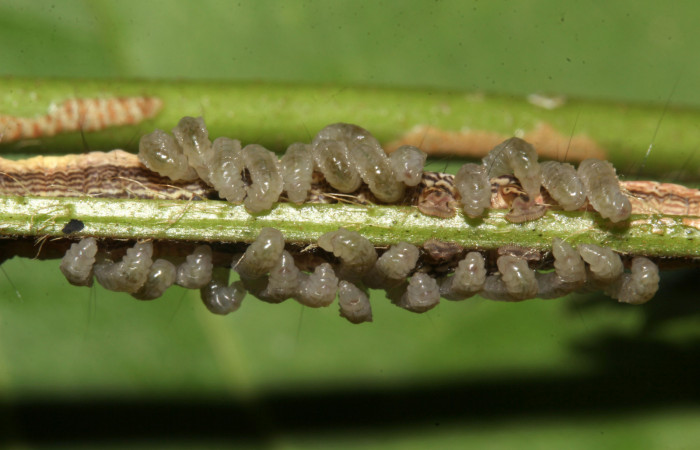
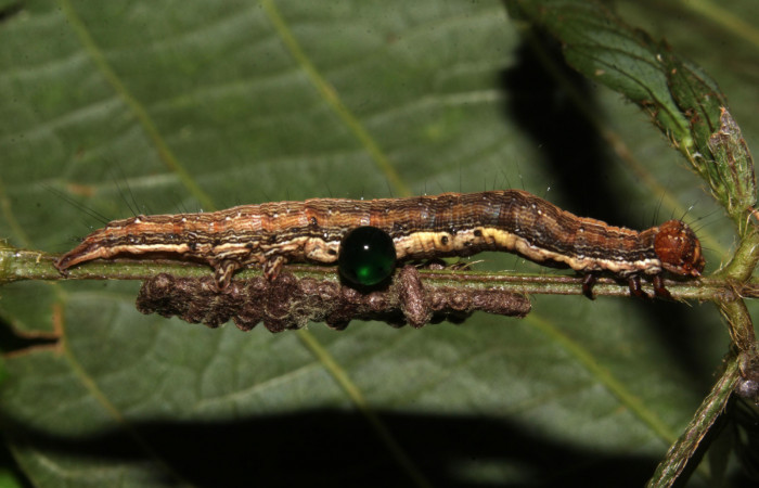
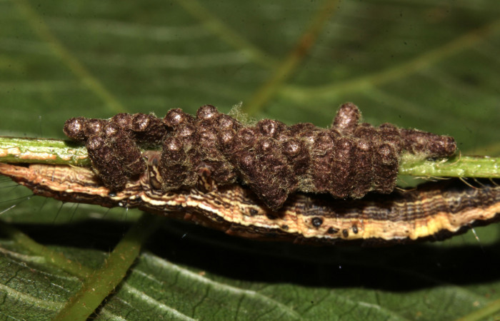
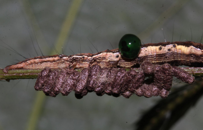
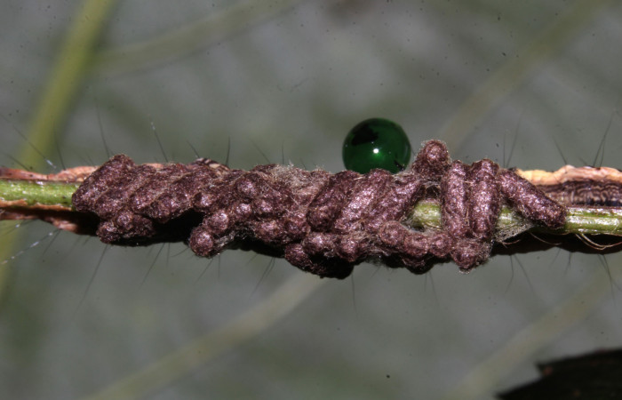
Parásitos de la familia Braconidae, saliendo de la larva (Fig. 23), estuvimos observando los parásitos mientras salían de la larva durante mas de 20 minutos hasta que hicieron los capullos (Fig. 17,18,19, 20, 21,22).
 Figura 17. Parásitos de la larva Lesmone formularis (Erebidae), estadio intermedio 33 mm. Larvas de Braconidae. Sector Pitilla. Foto: 09/Enero/2019. Voucher: 19-SRNP-30015-DHJ748564.jpg.
 Figura 18. Parásitos de la larva Lesmone formularis (Erebidae), estadio intermedio 33 mm. Larvas de Braconidae. Sector Pitilla. Foto: 09/Enero/2019. Voucher: 19-SRNP-30015-DHJ748566.jpg.
 Figura 19. Parásitos de la larva Lesmone formularis (Erebidae), estadio intermedio 33 mm. Larvas de Braconidae. Sector Pitilla. Foto: 09/Enero/2019. Voucher: 19-SRNP-30015-DHJ748565.jpg.
 Figura 20. Larva de Lesmone formularis (Erebidae), parásitada de Braconidae, estadio intermedio 33 mm. Larvas de Braconidae. Sector Pitilla. Foto: 09/Enero/2019. Voucher: 19-SRNP-30015-DHJ748558.jpg.
 Figura 21. Parásitos de la larva Lesmone formularis (Erebidae), estadio intermedio 33 mm. Larvas de Braconidae. Sector Pitilla. Foto: 09/Enero/2019. Voucher: 19-SRNP-30015-DHJ748560.jpg.
 Figura 22. Parásitos de la larva Lesmone formularis (Erebidae), estadio intermedio 33 mm. Larvas de Braconidae. Sector Pitilla. Foto: 09/Enero/2019. Voucher: 19-SRNP-30015-DHJ748561.jpg.
En estas imágenes también se puede apreciar una bolita verde (Fig. 23,24,25,26,) que corresponde a otro parásito de la familia Eulophidae del género Euplectrus sp (avispa) ella pega sus huevos directamente encima de la cutícula de la larva de la mariposa, y cuando sale la larvita del huevo, queda por fuera con su boca agarrando la cutícula, chupando la sangre de la larva de la mariposa. Cuando está totalmente desarrollada, la larva de la avispa construye su capullo afuera, con frecuencia pegado directamente al cadaver de la larva de la mariposa.
Tardaron como 25 minutos las larvas de Braconidae en salir de la larva Lesmone formularis (Erebidae) para formar los capullos.
 Figura 23. Larva de Lesmone formularis (Erebidae), estadio intermedio 33 mm. Capullos de Braconidae. Sector Pitilla. Foto: 09/Enero/2019. Voucher: 19-SRNP-30015-DHJ748569.jpg.
 Figura 24. Larva de Lesmone formularis (Erebidae), estadio intermedio 33 mm. Capullos de Braconidae. Sector Pitilla. Foto: 09/Enero/2019. Voucher: 19-SRNP-30015-DHJ748573.jpg.
 Figura 25. Larva de Lesmone formularis (Erebidae). estadio intermedio 33 mm. Capullos de Braconidae. Sector Pitilla. Foto: 09/Enero/2019. Voucher: 19-SRNP-30015-DHJ748570.jpg.
 Figura 26. Larva de Lesmone formularis (Erebidae). estadio intermedio 33 mm. Capullos de Braconidae. Sector Pitilla. Foto: 09/Enero/2019. Voucher: 19-SRNP-30015-DHJ748572.jpg.
Video de los parásitos de la familia Braconidae:
Larva Lesmone formularis (Erebidae), se observa encima las larvas de parásitos mientras salían, duraron mas de 20 minutos en salir, y luego quedaron pegadas a la larva. Se observa como éstas larvas parásitas empiezan a tejer su capullo fuera de la larva. Posteriormente saldrán las avispitas de la familia Braconidae.
Planta hospedera de Lesmone formularis (Erebidae), en Area de Conservación Guanacaste (ACG):
Sector Santa Rosa: con un elevación que va de los 290 a 305 metros, con 4 registros en Mimosa pigra (Fabaceae).
Sector Hrizontes: con un elevación de 180 metros, se han encontrado 2 registros, en Mimosa pigra (Fabaceae).
Sector Mundo Nuevo: con un elevación 325 a 480 metros , con 6 registros, 1 en Mimosa xanti (Fabaceae), 5 en Mimosa xantrocentra (Fabaceae).
Sector Pitilla: con un elevación que va de los 380 a 530 metros, con 33 registros, 31 en Mimosa quadrivalvis (Fabaceae), 1 en Zygia longifolia (Fabaceae), 1 Mimosa watsonii (Fabaceae).
Sector San Cristobal: con un elevación que va de los 460 a 645 metros, con 19 registros, 18 en Mimosa dormiens (Fabaceae), 1 en Mimosa guilandinae (Fabaceae).
Sector Rincon Rain Forest: con un elevación que va de los 123 a 491 metros, con 13 registros, 12 Mimosa dormiens (Fabaceae), 1 Mimosa pigra (Fabaceae).


